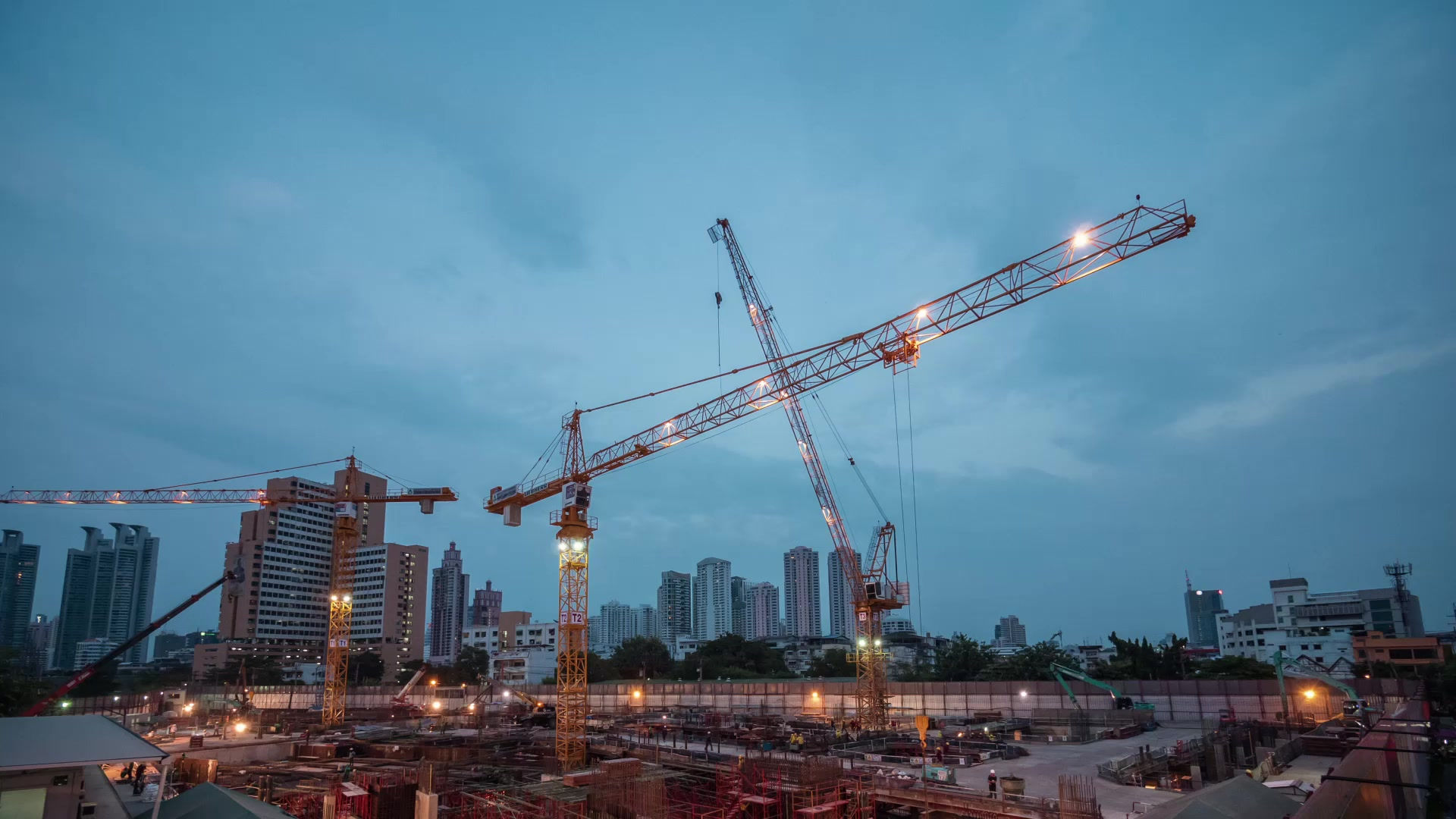
BS 6093 pdf free download: A guide to design of joints and jointing in building construction
- devyncictva7
- Aug 12, 2023
- 1 min read
We thank EXINI, Lund, Sweden, for the installation and guidance on the use of EXINI BoneBSI free of charge at the involved trial sites for the duration of the study. We thank Helle H. Eriksen, Aalborg University Hospital Statistical Consultancy Group, for assistance with the statistical analyses.
Markers internalized into animal cells appear sequentially in peripheral early endosomes, then in late endosomes, predominantly located in the perinuclear region, and eventually in lysosomes (review, Gruenberg and Howell, 1989; Kornfeld and Mellman, 1989; see Fig 1). Our major interest is to understand the mechanisms regulating membrane traffic in this pathway. For these studies, we use a cell-free assay measuring endocytic vesicle fusion. Avidin and biotinylated horseradish peroxidase (bHRP) are separately internalized by fluid phase endocytosis into two cell populations. After homogenization, endosomal fractions are prepared by immuno-isolation (Gruenberg and Howell, 1986, 1987; Gruenberg et al., 1989) or flotation on gradients (Tuomikoski et al., 1989; Bomsel et al., 1989; Gorvel et al., submitted). In the assay, avidin- and bHRP-labeled fractions are combined with cytosol and incubated at 37C in the presence of ATP and biotinylated insulin, as a scavenger (Braell, 1987; Gruenberg et al., 1989). When fusion occurs, a complex is formed between avidin and bHRP, which is then immuno-precipitated after detergent extraction. Fusion is quantified by measuring the enzymatic activity of bHRP in the complex. With this assay, we have reconstituted an early (Gruenberg et al., 1989) and a late (Bomsel et al., 1990) endocytic fusion event and we have shown that these reflect lateral interaction between early endosome elements and delivery to late endosomes, respectively.
Bs 6093 Pdf Free Download
2ff7e9595c

Comments